Prostate Imaging
Case Studies
Find your care
Our radiologists lead the way in prostate imaging. We offer the newest techniques to better detect and stage prostate cancer. Call 310-481-7545 to find out more about prostate imaging and treatment options.
1) Radiation Therapy Follow-Up
- 69 year-old, BPH gradual
- PSA rise 1.6 → 4.8 ng/dl
- Abnormal digital rectal exam → Biopsy: Gleason 5+4=9
- Read More >
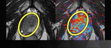
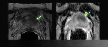
2) Small Volume, Low Grade Disease
- 60 year-old
- PSA 3.6
- 5 of 6 right biopsies GS3+3
- Read More >
3) Active Surveillance Example 1
- (Initial MRI order was for surgical planning)
- 65 year-old
- PSA 3 → 11 over 6 months
- Microfocus of Gleason 3+3
- Initial report : No posterior disease
- Targeted Biopsy consult prompted 2nd look
- Read More >
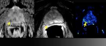
4) Example of Change to Nerve Sparing
- 59 year-old
- PSA 4.3
- standard biopsy: 3+3=6 but large volume left
- Read More >
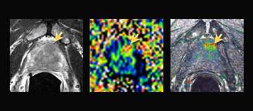
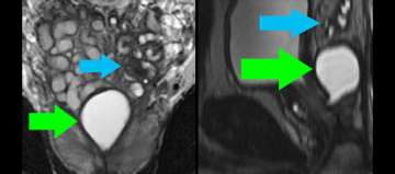
5) Extracapsular Extension and Seminal Vesicle Invasion by MRI + Spectroscopy and Pathology
- 59 year-old male
- PSA level: 10
- Clinical staging: T1C
- Blind systematic biopsy results: Gleason: 3+4=7 in the Left prostate
- Read More >
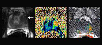
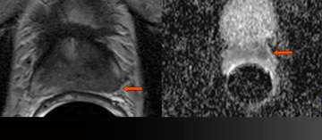
6) Dynamic Contrast-Enhancement for Biochemical Failure
- Elevated PSA after prostatectomy
- Read More >
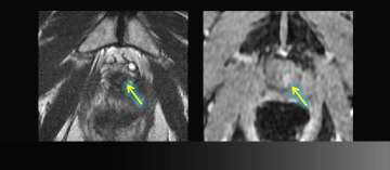
7) MR-Guided Targeted Biopsy
- 63 y/o, PSA 8.8 → 13.2 over 5 years
- All systematic biopsies negative
- Hypointense left anterior lesion with restricted diffusion is moderately suspicious, not in biopsy zone
- Read More >
8) Benign Disease (Infertility)
- Young man presents with infertility (azospermia)
- Workup and medical history otherwise noncontributory
- Muellerian duct remnant in central upper prostate may obstruct ejaculatory ducts
- Left seminal vesicles are atrophic
- Read More >