About Us
Find your care
Our radiologists lead the way in prostate imaging. We offer the newest techniques to better detect and stage prostate cancer. Call 310-481-7545 to find out more about prostate imaging and treatment options.

Our Mission
The UCLA Prostate Imaging and Treatment Program provides the most comprehensive and sophisticated imaging for all aspects of prostate disease. We aid referring health care providers with expertise in prostate cancer diagnosis, treatment, and subsequent follow up , as well as in the diagnosis of benign disease.
Our Program
Our program is supported by a multidisciplinary team of subspecialized experts including genitourinary radiology, medical physics, imaging diagnosis, imaging informatics, and MR spectroscopy. We use advanced MR imaging techniques such as perfusion, diffusion, diffusion tensor imaging and MR-ultrasound image fusion for biopsy guidance and direct MR guided biopsy. We work with our referring physicians to apply all relevant aspects of our expertise to address the specific concerns individual to each patient.
UCLA is Leading the Way in Prostate Imaging
Screening of Prostate Cancer
Currently, most clinicians rely strictly on clinical and laboratory parameters to guide decision making. Prostate cancer screening usually begins with a digital rectal exam (DRE) by a primary care physician or Urologist with a periodic evaluation of a patient's blood sample for prostate-specific antigen (PSA), a blood protein secreted by prostate cancer. In case of a palpable prostate nodule or elevated PSA, an ultrasound guided biopsy of the peripheral portion of the prostate is performed. The pathologist evaluates these samples under the microscope and decides whether the features of the prostate glands suggest the presence of cancer. If present, the cancer severity is graded using the Gleason Score.
New Techniques to Better Detect & Stage Prostate Cancer
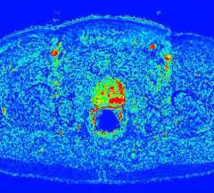
In addition to MRI and MRSI, newer techniques such as MR diffusion and MR perfusion imaging are being routinely used to better detect and stage prostate cancer to assist surgeons in planning minimally invasive surgery.
Diffusion imaging is a much faster technique which, although still not as finely detailed as conventional MRI, provides information on the degree of cellular crowding, which is worse where cancer cells are rapidly turning over. Perfusion imaging has nearly the same fine detail as normal MRI, but provides a map of blood flow, which is increased in cancer cells. This last technique is just now exiting the realm of experimental use and is being gingerly applied to patient diagnosis and treatment planning (fig.3).
These advanced preoperative imaging techniques enabled surgeons to more accurate decided when to remove or spare the delicate neurovascular bundles in different cases. These decisions do significantly affect patients' quality of life after surgery since urinary continence and sexual potency may be affected.
In some cases, a blood test may suggest cancer but the biopsies fail to localize it, and the surgeon may not be able to feel the cancer with the DRE. In these cases UCLA can provide magnetic resonance imaging with these new and established techniques to help find cancer and guide future biopsies.