• UCLA Health
  • myUCLAhealth
  • School of Medicine
UCLA Prostate Imaging and Treatment

UCLA Prostate Imaging and Treatment

UCLA Prostate Imaging and Treatment
  • Back to Main Radiology
  • About Us
    • Case Studies
    • Clinical Trials
    • CME Course
    • Research Publications
  • Conditions Treated
    • Prostate Cancer
    • Benign Prostatic Hyperplasia
  • Treatments & Procedures
    • Imaging and Biopsy Services
    • Prostate Artery Embolization
    • TULSA
  • Schedule an Appointment
  • Our Expert Team
  • UCLA Health
  • myUCLAhealth
  • School of Medicine

UCLA Prostate Imaging and Treatment

About Us

About Us

About Us

  • Case Studies
    • Radiation Therapy
    • Small Volume Low Grade Disease
    • Active Surveillance
    • Example of Change to Nerve Sparing
    • Extracapsular Extension and Seminal Vesicle Invasion
    • Dynamic Contrast-Enhancement for Biochemical Failure
    • MR-Guided Targeted Biopsy
    • Benign Disease (Infertility)
  • Clinical Trials
  • CME Course
  • Research Publications
  • Case Studies
  • Clinical Trials
  • CME Course
  • Research Publications
  • Radiation Therapy
  • Small Volume Low Grade Disease
  • Active Surveillance
  • Example of Change to Nerve Sparing
  • Extracapsular Extension and Seminal Vesicle Invasion
  • Dynamic Contrast-Enhancement for Biochemical Failure
  • MR-Guided Targeted Biopsy
  • Benign Disease (Infertility)
  1. Home
  2. UCLA Prostate Imaging and Treatment
  3. About Us
  4. Case Studies
  5. MR-Guided Targeted Biopsy

MR-Guided Targeted Biopsy

Share this

History

  • 63 y/o, PSA 8.8 → 13.2 over 5 years
  • All systematic biopsies negative
  • Hypointense left anterior lesion with restricted diffusion is moderately suspicious, not in biopsy zone

Imaging


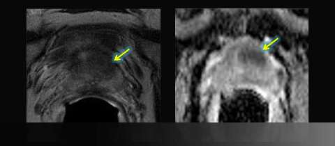
LEFT: Axial T2-weighted image: asymmetric anterior low signal
RIGHT: Apparent diffusion coefficient (ADC) map: focal restricted diffusion


Oblique Axial T2-weighted image
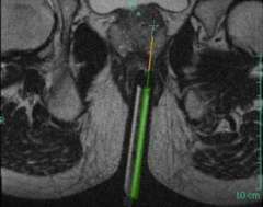
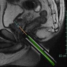
LEFT: Oblique Axial T2-weighted image from in-bore MR-guided targeting
Corresponding sagittal T2-weighted image
RIGHT: Corresponding sagittal T2-weighted image from in-bore MR-guided targeting


Results

  • Gleason grade 3+3=6/10
  • All biopsies over 5 mm and over 50% of core

Advantage: UCLA Prostate MRI

Suspicious findings on diagnostic multiparametric prostate MRI can be targeted for direct in-bore MRI-guided biopsy or MRI-ultrasound fusion targeted biopsy

Like Us on Facebook Follow Us on Twitter Subscribe to Our Videos on YouTube Follow us on Instagram Connect with Us on LinkedIn Follow us on Pinterest
UCLA Health hospitals ranked best hospitals by U.S. News & World Report
  • UCLA Health
  • Find a Doctor
  • School of Medicine
  • School of Nursing
  • UCLA Campus
  • Directory
  • Newsroom
  • Subscribe
  • Patient Stories
  • Giving
  • Careers
  • Volunteer
  • International Services
  • Privacy Practices
  • Nondiscrimination
  • Billing
  • Health Plans
  • Emergency
  • Report Broken Links
  • Terms of Use
  • 1-310-825-2631
  • Maps & Directions
  • Contact Us
  • Your Feedback
  • Report Misconduct
  • Get Social
  • Sitemap
Like Us on Facebook Follow Us on Twitter Subscribe to Our Videos on YouTube Follow us on Instagram Connect with Us on LinkedIn Follow us on Pinterest

Sign in to myUCLAhealth

Learn more about myUCLAhealth