• UCLA Health
  • myUCLAhealth
  • School of Medicine
UCLA Prostate Imaging and Treatment

UCLA Prostate Imaging and Treatment

UCLA Prostate Imaging and Treatment
  • Back to Main Radiology
  • About Us
    • Case Studies
    • Clinical Trials
    • CME Course
    • Research Publications
  • Conditions Treated
    • Prostate Cancer
    • Benign Prostatic Hyperplasia
  • Treatments & Procedures
    • Imaging and Biopsy Services
    • Prostate Artery Embolization
    • TULSA
  • Schedule an Appointment
  • Our Expert Team
  • UCLA Health
  • myUCLAhealth
  • School of Medicine

UCLA Prostate Imaging and Treatment

About Us

About Us

About Us

  • Case Studies
    • Radiation Therapy
    • Small Volume Low Grade Disease
    • Active Surveillance
    • Example of Change to Nerve Sparing
    • Extracapsular Extension and Seminal Vesicle Invasion
    • Dynamic Contrast-Enhancement for Biochemical Failure
    • MR-Guided Targeted Biopsy
    • Benign Disease (Infertility)
  • Clinical Trials
  • CME Course
  • Research Publications
  • Case Studies
  • Clinical Trials
  • CME Course
  • Research Publications
  • Radiation Therapy
  • Small Volume Low Grade Disease
  • Active Surveillance
  • Example of Change to Nerve Sparing
  • Extracapsular Extension and Seminal Vesicle Invasion
  • Dynamic Contrast-Enhancement for Biochemical Failure
  • MR-Guided Targeted Biopsy
  • Benign Disease (Infertility)
  1. Home
  2. UCLA Prostate Imaging and Treatment
  3. About Us
  4. Case Studies
  5. Radiation Therapy

Radiation Therapy

Share this

History

  • 69 year-old, BPH gradual
  • PSA rise 1.6 → 4.8 ng/dl
  • Abnormal digital rectal exam → Biopsy: Gleason 5+4=9

Imaging


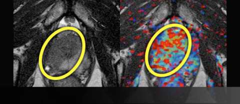
LEFT: T2-weighted image prior to treatment shows large, low-signal mass (yellow oval) on the right crossing midline anteriorly
RIGHT: Color perfusion map shows correspondingly increased perfusion (yellow-green pixels) with washout (red pixels)
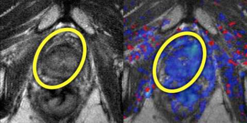


Left: T2-weighted image after treatment shows normalization of T2 signal with reappearance of the normal peripheral-transitional zone border.
Right: Color perfusion map shows correspondingly decreased perfusion (blue pixels) consistent with response to treatment.


Example Report (Prior to Therapy) test

* Findings

No suspicious lymph nodes or bone lesions
Prostate measures 58 gm with mild prostatic hyperplasia
A single suspicious area is identified:

Location Bilateral anterior & right posterior
Size 2.8 cm
Capsule Involvement posterolaterally
T2 Asymmetric, ill-defined
Perfusion Abnormal
Overall suspicion High (4/5)

Seminal vesicles and neurovascular bundles appear normal

* Impression

  1. Technical quality: excellent
  2. Large right lesion crosses midline anteriorly and extends to capsule
  3. Organ-confined disease - no suspicious lymph nodes or bone lesions

Advantage: UCLA Prostate MRI

  • Although PSA remains the mainstay of treatment monitoring, MRI can add value when there is concern for residual disease
  • Removable table (GE HDxt or Siemens Skyra) allows for imaging after external patient mold is applied for registration with radiation planning.
Like Us on Facebook Follow Us on Twitter Subscribe to Our Videos on YouTube Follow us on Instagram Connect with Us on LinkedIn Follow us on Pinterest
UCLA Health hospitals ranked best hospitals by U.S. News & World Report
  • UCLA Health
  • Find a Doctor
  • School of Medicine
  • School of Nursing
  • UCLA Campus
  • Directory
  • Newsroom
  • Subscribe
  • Patient Stories
  • Giving
  • Careers
  • Volunteer
  • International Services
  • Privacy Practices
  • Nondiscrimination
  • Billing
  • Health Plans
  • Emergency
  • Report Broken Links
  • Terms of Use
  • 1-310-825-2631
  • Maps & Directions
  • Contact Us
  • Your Feedback
  • Report Misconduct
  • Get Social
  • Sitemap
Like Us on Facebook Follow Us on Twitter Subscribe to Our Videos on YouTube Follow us on Instagram Connect with Us on LinkedIn Follow us on Pinterest

Sign in to myUCLAhealth

Learn more about myUCLAhealth