Parathyroid 4D-CT Scan
Find Your Care
We deliver effective, minimally invasive treatments in a caring environment. Call 310-267-7838 to connect with an expert in endocrine surgery.
Four-dimensional parathyroid CT (4D-CT) is a specialized imaging study that was first introduced in 2006 for the purpose of identifying abnormal parathyroid glands in the planning of parathyroid surgery.
The patient is given intravenous contrast, which is an iodine-based dye that enhances the quality of imaging. Images are then acquired with the CT scanner at very specific times when the contrast is maximally taken up by the parathyroid glands. (The "fourth dimension" is time.)

Parathyroid 4D-CT Scan
Parathyroid surgery can be done through an incision measuring 1.5-2.0 cm in length.
Patient Experience Video.
Parathyroid 4D-CT Scan
Using 4D-CT imaging, surgeons locate abnormal parathyroid glands before operating.
Patient Experience Video.
Advantages of 4D-CT
Parathyroid 4D-CT represents the latest technology in parathyroid imaging. The resolution of 4D-CT imaging is higher than that of any other type of parathyroid scan. In fact, we have identified abnormal parathyroid glands as small as 1 mm x 6 mm which we have then gone on to remove, resulting in successful, curative parathyroid surgery.
Parathyroid 4D-CT is particularly useful when other imaging studies have been negative (failed to show the abnormal parathyroid gland) or when the anatomy of the neck has been distorted by prior surgery or co-existent thyroid disease. In our experience at UCLA of reading several hundred scans per year, we have been able to detect at least one abnormal parathyroid gland in 99% of cases. In other words, parathyroid 4D-CT is a highly sensitive scan.
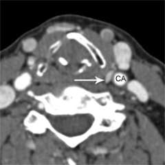
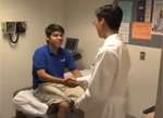
Figure 1a. Parathyroid 4D-CT showing a parathyroid adenoma in an abnormal position within the carotid sheath, missed during a prior surgery. CA= Carotid artery.
How is 4D-CT different from a regular CT scan?
This is a very common question that we get from patients, doctors, and insurance companies. Parathyroid 4D-CT is not the same as a regular CT scan. There are several key differences:
- Patient positioning. Patients are positioned in an optimized way that minimizes interference from other structures such as the teeth, jaws, dental fillings and/or other dental appliances, the shoulders, and collar bones.
- Amount and timing of intravenous contrast. A specific volume of intravenous contrast is given at a specific rate, at a specific time.
- Timing of image acquisition and radiation dosage. Imaging is acquired in three phases: pre-contrast, arterial phase, and venous phase. The timing of these phases is critical, and allows us to tell apart parathyroid tissue from other adjacent tissues.
- Interpretation of the scan by an experienced radiologist. All of our parathyroid 4D-CT scans are supervised and interpreted by a single experienced radiologist. (See Who interprets the results? below)
In a word, parathyroid 4D-CT is different from a regular CT scan because we have spent considerable effort optimizing 4D-CT for our patients with challenging parathyroid disease. This process of continuous quality improvement took several years: one year to optimize the scanning technique (2011), another year to validate the results in comparison to actual surgical findings in a standard patient population (2012), and a third year (2013) to validate the results in a more challenging patient population (principally patients who had had failed parathyroid surgery elsewhere who came to us for re-do surgery). If you have had a regular CT scan of the neck performed elsewhere, it generally cannot be used to plan your parathyroid surgery. Please have your imaging done at UCLA on our equipment if you would like to have surgery here.
Figure 1b. Parathyroid 4D-CT showing a parathyroid adenoma in an abnormal position within the carotid sheath, missed during a prior surgery. CA= Carotid artery.
Who needs a parathyroid 4D-CT?
Parathyroid 4D-CT is most useful when other imaging studies are negative, and in particular in re-operative cases. Ultrasound and sestamibi are still the first-line imaging studies used to detect abnormal parathyroid glands. If these tests are able to localize the abnormal gland, there is generally no need for additional imaging. If both your ultrasound and sestamibi are negative, a parathyroid 4D-CT will usually show the abnormal gland or suggest that multiple glands are abnormal. Many of our patients and referring doctors like this, because it provides an increased sense of security going in to surgery. If desired, parathyroid 4D-CT can be performed without first obtaining a sestamibi scan.
The most important role of parathyroid 4D-CT is in the management of re-operative cases (patients with a history of prior failed parathyroid surgery). In these patients, the fine resolution provided by parathyroid 4D-CT helps us to minimize dissection during surgery, simultaneously increasing the likelihood of cure while reducing the risk of surgical complications. Our research has shown that parathyroid 4D-CT has made re-operative surgery both faster and more successful, particularly in very challenging cases where multiple abnormal parathyroid glands are causing the patient's disease. Indeed, a number of patients we have recently cured with successful re-operation would have previously been considered incurable (ineligible for surgery) prior to the advent of parathyroid 4D-CT. So, if you have been told that you are ineligible for re-operative parathyroid surgery because of negative imaging, obtaining a parathyroid 4D-CT would be a good next step.
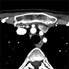
Figure 2. Parathyroid 4D-CT showing a parathyroid adenoma in an abnormal position within the mediastinum.

Figure 3. Parathyroid 4D-CT showing a parathyroid adenoma in an abnormal position within the mediastinum.
How do I get a parathyroid 4D-CT?
A parathyroid 4D-CT needs to be performed at an experienced, high-volume center. Currently there are approximately 10 centers in the United States, including UCLA, that have a dedicated parathyroid 4D-CT imaging program and have published their results in the medical literature. If you are interested in having a parathyroid 4D-CT performed at UCLA, you or your doctor may call UCLA Endocrine Surgery at (310) 206-0585, or the UCLA Radiology Department at (310) 301-6800.
What is the procedure for getting scanned?
A parathyroid CT scan takes about 5 minutes to perform, though you should set aside about one hour, to include registration and preparation time. It can usually be scheduled within 1-2 days of us ordering it. The images are immediately available, and it is our practice to review the images in person with the patient and patient's family to maximize their understanding of the findings.
Who interprets the results?
At UCLA, Dr. Ali Sepahdari oversees the parathyroid 4D-CT imaging program and interprets all of our studies. Dr. Sepahdari is an experienced Head and Neck Radiologist who has worked closely with us to continuously improve the quality of our scans.
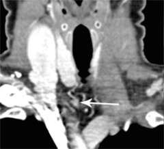
Figure 4a. Parathyroid 4D-CT showing multiple gland hyperplasia in the same patient, missed in a prior surgery. Enlarged right inferior parathyroid gland, below the right thyroid lobe.
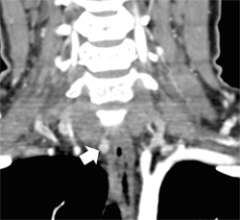
Figure 4b. Parathyroid 4D-CT showing multiple gland hyperplasia in the same patient, missed in a prior surgery. Enlarged right superior parathyroid gland, next to the esophagus.
How much radiation does it involve?
As more patients get CT scans for various different reasons, we are all concerned about the risks of radiation exposure. We have worked to minimize the amount of radiation used during parathyroid 4D-CT and the dosage we use is among the lowest of all centers that perform this study.
Will it be covered by insurance?
In most cases, yes. For patients who are out of network, we will work with you to achieve an affordable rate.
My doctor says I can't have a CT scan with contrast because I have kidney problems. Now what?
We do need to be careful with administering intravenous contrast to patients with kidney disease (your doctor may tell you about an elevated creatinine level in the blood, which is a measure of kidney function). The good news is that we are able to take extra precautions that make the study safe for most patients with mild to moderate kidney disease. If your kidney function is below a certain level, we may need to develop a more creative solution for you. Please contact us for more information.
References
- Cham S, Sepahdari AR, Hall KE, Yeh MW, Harari A. Dynamic Parathyroid Computed Tomography (4DCT) Facilitates Reoperative Parathyroidectomy and Enables Cure of Missed Hyperplasia. Ann Surg Onc, in press.
- Sepahdari AR, Bahl M, Harari A, Kim HJ, Yeh MW, Hoang JK. Predictors of Multigland Disease in Primary Hyperparathyroidism: A Scoring System with 4D-CT Imaging and Biochemical Markers. AJNR Am J Neuroradiol. 2015 Jan 2.
- Rodgers SE, Hunter GJ, Hamberg LM, et al. Improved preoperative planning for directed parathyroidectomy with 4-dimensional computed tomography. Surgery 2006;140(6):932–940.
- Lubitz CC, Hunter GJ, Hamberg LM, et al. Accuracy of 4-dimensional computed tomography in poorly localized patients with primary hyperparathyroidism. Surgery. 2010 Dec;148(6):1129-37.
- Mortenson MM, Evans DB, Lee JE, et al. Parathyroid exploration in the reoperative neck: improved preoperative localization with 4D-computed tomography. J Am Coll Surg 2008;206(5):888–895.
- Kelly HR, Hamberg LM, Hunter GJ. 4D-CT for preoperative localization of abnormal parathyroid glands in patients with hyperparathyroidism: accuracy and ability to stratify patients by unilateral versus bilateral disease in surgery-naive and re-exploration patients. AJNR Am J Neuroradiol. 2014 Jan;35(1):176-81.
- Madorin CA, Owen R, Coakley B, et al. Comparison of radiation exposure and cost between dynamic computed tomography and sestamibi scintigraphy for preoperative localization of parathyroid lesions. JAMA Surg. 2013 Jun;148(6):500-3.
- Mahajan A, Starker LF, Ghita M, et al. Parathyroid four-dimensional computed tomography: evaluation of radiation dose exposure during preoperative localization of parathyroid tumors in primary hyperparathyroidism. World J Surg 2012;36(6):1335–1339.