As organoids become an increasingly important tool for studying human biology and disease, UCLA researchers are working across disciplines to expand how these three-dimensional laboratory models can be used to answer complex questions about cancer, immune function and other areas of health.
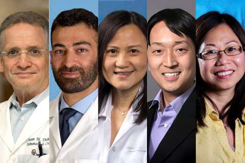
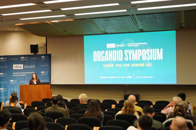
The effort was highlighted at the UCLA Health Jonsson Comprehensive Cancer Center’s inaugural Organoid Research Symposium, held in partnership with the UCLA Broad Stem Cell Research Center, the David Geffen School of Medicine at UCLA and the UCLA Division of Life Sciences. The symposium was co-organized by Brigitte Gomperts, MD, and Gay Crooks, MD, who co-lead the Cancer Center’s Stem Cell Biology Research Program.
The event brought together researchers from cancer biology, stem cell biology, bioengineering, computational science, imaging and other fields to share how they are using organoids and discuss ways to make the technology more accessible, reproducible and scalable across campus.
Organoids offer researchers a way to recreate aspects of human biology that can be difficult to capture in traditional cell cultures or animal models. Depending on how they are designed, organoids can help researchers investigate how tumors evolve, how cancer cells interact with surrounding tissues, why treatments stop working and even how human tissues and organs develop.
Despite their potential, researchers still face challenges in developing and using organoid models. The models can be difficult to reproduce consistently, protocols vary among laboratories and researchers may lack access to the specialized expertise, equipment or models needed to use them effectively.
“Our goal today is to start the process of creating an organoid research community at UCLA that overcomes these practical hurdles to advance the science,” said Dr. Gomperts. “No single lab holds all the answers. We need to be able to communicate across all these fields.”
The symposium also featured a keynote address from David Tuveson, MD, PhD, director of the Cold Spring Harbor Laboratory Cancer Center and a leader in developing organoid models of pancreatic cancer. Drawing on his experience building an organoid research program, Tuveson emphasized that realizing the potential of the technology requires sustained resources, expertise and collaboration.
“It’s far more than growing conventional cell lines,” Tuveson said. “But it can be accomplished if there’s a collective commitment.”
Using organoids to better understand cancer
The symposium showcased the many ways UCLA researchers are using organoids to study cancer and human biology.
Mingxia Gu, MD, PhD, professor of anesthesiology and perioperative medicine, is developing tumor organoids that incorporate specialized blood and lymphatic vessels to create more complete models of the tumor microenvironment.
These models could allow researchers to directly observe how immune cells travel through blood vessels and enter tumors, determine whether cancer drugs can effectively cross the tumor vasculature to reach cancer cells, and study how tumor cells enter lymphatic vessels and begin to spread. By recreating these processes in a human organoid system, the approach could help explain why some tumors resist treatment, identify ways to improve drug delivery and immune-cell access, and provide a platform for testing therapies designed to prevent cancer from spreading.
Other researchers are using organoids to understand how cancers change over time and adapt to treatment.
Andrew Goldstein, PhD, professor of molecular, cell and developmental biology and urology, and his team developed a human prostate cancer organoid model that recreates an earlier, hormone-sensitive stage of the disease that has been difficult to study using existing models. By introducing cancer-driving genetic changes in different sequences, researchers can investigate how those changes affect response to hormone therapy and how prostate cancer evolves into treatment-resistant disease.
To understand glioblastoma, one of the most difficult brain cancers to treat, David Nathanson, PhD, professor of molecular and medical pharmacology, and colleagues are using human brain organoids developed by Aparna Bhaduri, PhD, assistant professor of medicine and biological chemistry, in which glioma cells are co-cultured with human brain organoids, to study how tumor cells adapt to treatment. Their work suggests glioblastoma cells can shift between different cellular states when exposed to targeted therapy, potentially helping tumors continue growing despite treatment. By uncovering a common signaling mechanism that allows tumor cells to make these adaptations, the team also identified a potential therapeutic vulnerability, opening a new avenue for preventing treatment resistance.
Harley Kornblum, MD, PhD, professor of psychiatry, pharmacology and pediatrics and Director of the UCLA Intellectual and Developmental Disabilities Research Center, is using human gliomasphere models that he developed as well as the mixed human glioma-brain organoid system used by Dr. Nathanson to study another aspect of tumor behavior. His team's research suggests glioblastoma cells can transfer small pieces of cancer-associated DNA, known as extrachromosomal DNA, to other tumor cells and potentially to neighboring noncancerous cells. Researchers are investigating whether this exchange helps tumors evolve or changes surrounding cells in ways that support tumor growth.
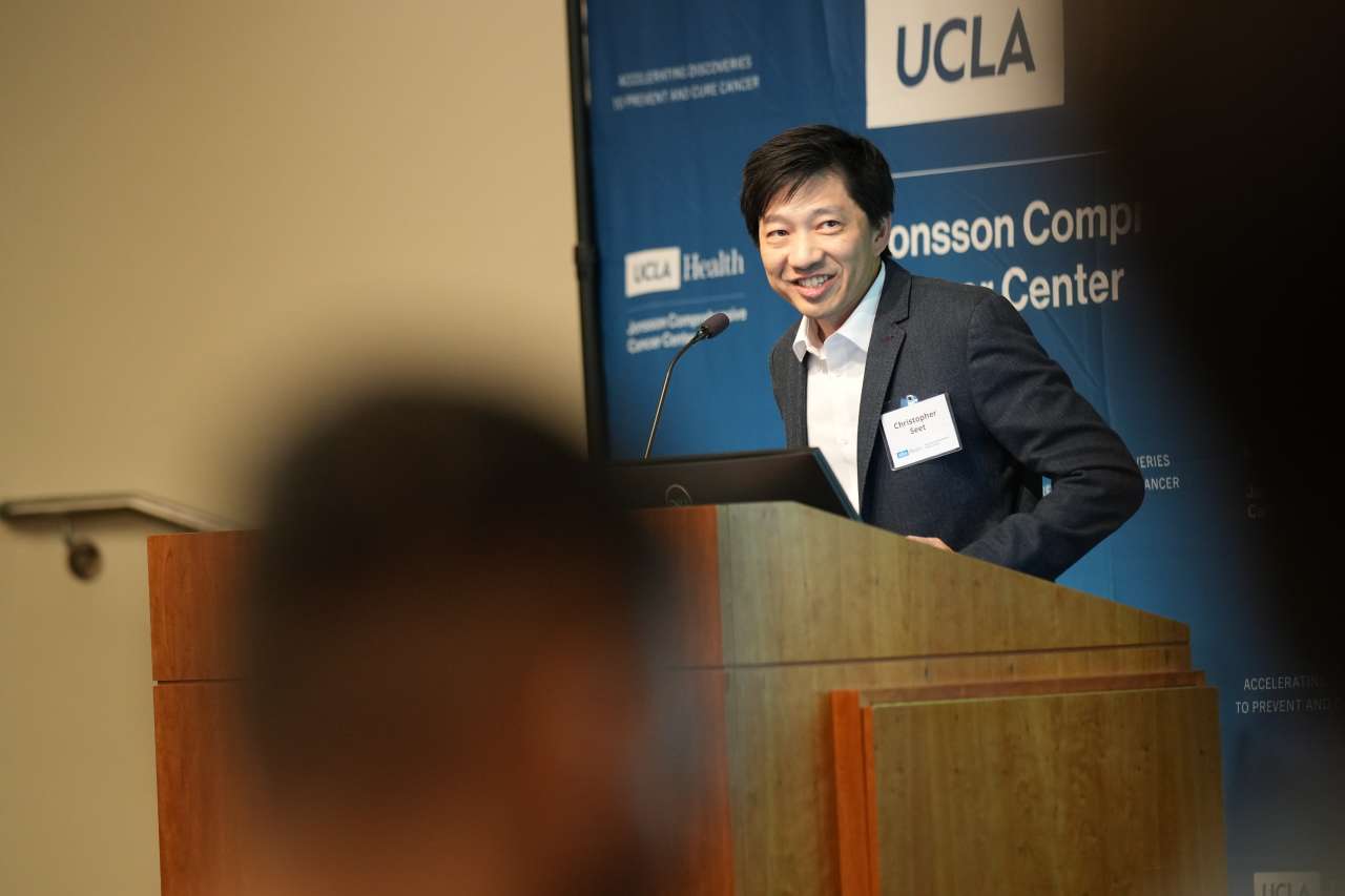
Organoids can also provide new ways to study and develop cancer immunotherapies. Christopher Seet, MD, PhD, assistant professor of medicine, and Dr. Crooks developed an artificial thymic organoid that recreates key aspects of the thymus, the organ where T cells develop.
The system allows researchers to study in detail how T cells develop from stem cells, and test genetic engineering approaches that could be used to make T cells more effective in treating cancer. Seet's team has used the approach to generate CAR T cells engineered to recognize cancer targets and is studying how these stem-cell-derived cells compare with conventional CAR T cells from the blood.
Making organoid research more reproducible
As researchers develop increasingly complex organoid models, the symposium also highlighted the importance of making them more consistent and scalable.
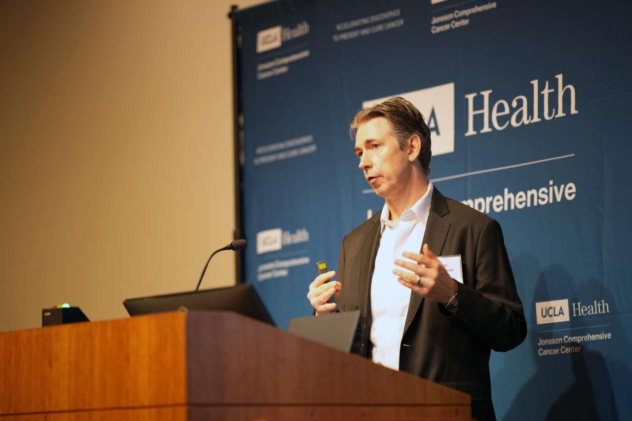
Robert Damoiseaux, PhD, professor of molecular and medical pharmacology and bioengineering and director of UCLA's Molecular Screening Shared Resource at the California NanoSystems Institute, presented automated technologies that reduce the variability introduced when organoids and stem cells are cultured and analyzed by hand.
The platform, acquired through an NIH shared instrumentation award, combines automated cell culture, scheduled imaging and machine learning to perform more than monitoring: image analysis decides what happens next — when to feed, passage or image — and every intervention is logged against the image that prompted it. In a published head-to-head comparison, cultures handled by hand showed differences in pluripotency markers between two media that disappeared when the same experiment was run on the platform, indicating the difference came from handling rather than biology.
The system is in production and open to investigators at UCLA and beyond and is supporting work ranging from tumor organoid growth to a multi-donor screen of roughly 4,700 environmental compounds. Damoiseaux's team is now working with UCLA Pathology to develop patient-derived organoids for use by investigators across the campus.
Another challenge is determining how closely an organoid represents the tumor from which it was created.
Paul Spellman, PhD, professor of medicine and human genetics, discussed his team's work as part of the GDAN consortium evaluating models developed through the Human Cancer Models Initiative, a large collection of patient-derived cancer models that include organoids from a range of tumor types.
By comparing the models with the tumors from which they were derived, researchers found that most retained key genetic and molecular characteristics of the original cancers. More than 90% of the tumor-model pairs with sufficient data showed high agreement across the measurements examined.
The collection gives researchers access to hundreds of molecularly characterized cancer models, allowing them to identify models with genetic features relevant to the cancers they want to study.
Building connections across UCLA
While the research presented at the symposium spanned different diseases, technologies and scientific questions, a common theme was the value of bringing together expertise from across disciplines.
For Dr. Gomperts and Dr. Crooks, the inaugural symposium was an opportunity to begin building those connections at UCLA and encourage researchers who may approach organoids from very different perspectives to learn from one another.
“The symposium displayed just some of the tremendous activity in organoid technology already at UCLA, and the large number of investigators and trainees who attended has shown us all how important it will now be to build a collaborative network on campus for this exciting technology,” said Crooks.