Aneurysm and Dissection
Find your care
Our board-certified physicians diagnose and treat many vascular conditions. Call 310-481-7545 to learn more about interventional radiology services.
What is an aneurysm or dissection?
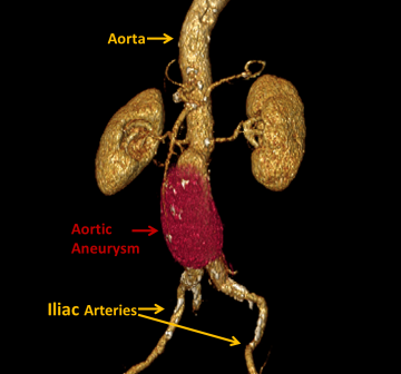
An aneurysm is a weakening of a vessel wall with resultant dilation of a segment of the vessel. As blood continues to flow in an aneurysm, the pressure on the weakened wall can cause the aneurysm to expand like a balloon. Aneurysms can occur in any vessel, most notably in the brain, heart, thoracic aorta, and abdominal aorta.
A dissection is a tear of the inside layer of a blood vessel wall that allows blood to flow between the layers that make up the vessel wall and separate these layers. This can compromise blood flow in the main portion of the vessel and result in loss of blood flow to critical organs. Dissections can also occur in any vessel, but most notably in the thoracic or abdominal aorta.
Aneurysms and dissections can result in areas of turbulent blood flow which can lead to the formation of blood clots, or thrombi. Blood clots that break off, called emboli, can cause damage if they lodge in the brain, heart, or other organs, resulting in reduced blood flow (ischemia) to the organ. An enlarged vessel (from either a dissection or an aneurysm) can also compress surrounding nerves and cause pain. When an aneurysm or dissection grows larger, the risk of blood vessel rupture increases. A rupture can lead to rapid bleeding and death if not treated.
Risk Factors
- History of aneurysm
- Increasing age
- Family history
- Genetic conditions such as Marfan Syndrome and Turner Syndrome
- Connective tissue disorders such as Ehler-Danlos Syndrome and Autosomal Dominant Polycystic Kidney Disease
- Certain infections such as syphilis
- Obesity
- Hypertension
- Atherosclerosis
- Alcoholism
- Smoking
Symptoms
Common symptoms of aneurysms and dissections are listed below. Intracranial (brain) aneurysms are not discussed here.
Unruptured Aneurysms
Symptoms for unruptured aneurysms vary depending on the location of the aneurysm, or they could be asymptomatic, depending on the size of the aneurysm. Common variations are listed below.
Thoracic aortic aneurysm
- Jaw, neck, chest, or upper back pain
- Shortness of breath
- Difficulty swallowing
- Hoarseness
Abdominal aortic aneurysm
- Abdominal or lower back pain
- Feeling a pulsation in the abdomen
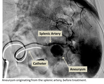
Visceral aneurysms
Visceral aneurysms are aneurysms in arteries that supply internal organs, such as the spleen, liver, intestines, and kidneys. Symptoms will vary depending on the organ the affected artery supplies.
Ruptured aneurysms
A ruptured aneurysm of a blood vessel such as the aorta can cause rapid large volume blood loss, causing hypovolemic shock as there is not enough blood pressure to deliver blood to the organs. Ruptured visceral aneurysms can cause similar symptoms. If not treated quickly, death can result.
- Sudden, severe pain upon rupture
- Abdominal swelling
- Pale skin
- Excessive thirst
- Nausea and vomiting
- Dizziness
- Rapid heart rate
- Hypovolemic shock
Dissection
- Sharp tearing or stabbing pain
- Aortic dissection usually includes severe chest pain that radiates to the back
- Difficulty swallowing
- Nausea and vomiting
- Shortness of breath
- Pale, clammy skin
- Dizziness
- Abdominal pain
Diagnosis
Some aneurysm cases may be diagnosed from a physical exam by your healthcare provider. The following tests are typically performed to determine if an aneurysm or dissection is present:
- CT or MRI with contrast
- Angiography (X-ray with contrast agent to visualize blood vessels)
- Doppler ultrasound
Treatments
Endovascular stent grafting (EVAR, or endovascular aneurysm repair)
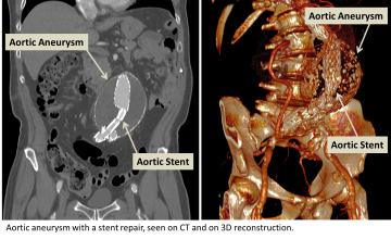
Endovascular stent grafting is a procedure that is performed to treat aneurysm or and dissection. A stent graft is an expandable vascular conduit which is placed into the diseased segment of vessel thereby reinforcing the vessel wall and reducing the potential of rupture or progression of disease.
- The procedure is performed by using a need to access a vessel in the arm or leg and placing wire into the vessel. The wire is navigated into the aneurysmal or dissected vessel using a fluroscope (x-ray machine) to guide the navigation.
- Once the wire is properly placed, an appropriate stent graft is selected based on many factors including the size of the vessel, the types of branches that come from the diseased vessel, and the type of aneursym or dissection being treated. The collapsed stent graft is fed into the target vessel using the wire and fluoroscope as a guide.
- Once the stent graft is appropriately positioned, a balloon is placed over the wire and into the center to the collapsed stent and used to expand the stent to seat it against the vessel wall.
- Contrast may be injected into the vessel to test the fit and seating of the stent graft. Once a satisfactory placement has been made, the wire and balloon are removed from the body and the puncture made in the arm or leg is sealed. The patient is then taken to the recovery area for post-procedural observation.
Aneurysm embolization
If the aneurysm is in a suitable location in the body and has specific dimensions, embolization is another treatment that may be considered.
- Aneurysm embolization is also performed by accessing the diseased blood vessel through a needle puncture in the arm or the leg. A wire is navigated into the aneurysmal vessel using a fluroscope (x-ray machine) to guide the navigation.
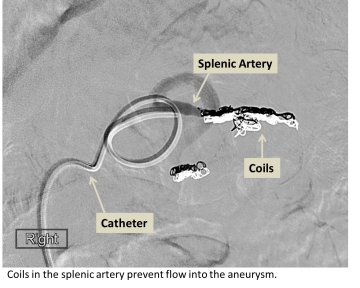
- Once the vascular tree has been accessed, a catheter is placed over the wire and used to navigate to the aneurysm. Embolization coils are then placed through the catheter into the aneurysm sac filling the sac and slowing blood flow into the sac. Once blood flow within the sac becomes slow enough, the body’s natural clotting cascade causes clot formation within the sac and blood flow within the sac is stopped.
- Contrast may be injected into the vessel to test that the coil pack is properly seated and blood flow into the aneurysm sac is slow enough. Once a satisfactory placement has been made, the wire and catheter is removed from the body and the puncture made in the arm or leg is sealed. The patient is then taken to the recovery area for post-procedural observation.
For More Information:
For more information or to schedule an appointment with one of our IR physicians, please call 310-481-7545.