Basics of Breast Ultrasound
by: Justin Sun, MD, Fadi Dahoud, and Olivia Linden, MD
Ultrasound is an imaging modality with broad applications for the characterization of soft tissue, including the breast. An electrical current is applied to piezoelectric crystals within a transducer, which in turn emit sound waves. These sound waves are transmitted through, and reflected off, tissues in a manner dictated by physical properties and interfaces. These reflected sound waves are carried back to the transducer, which translates them into a final image.
Indications for breast ultrasound are varied and range from breast cancer screening to advanced diagnostic evaluation. Some of these indications include: breast cancer screening for women with dense breasts (in addition to mammography), work up of symptomatic patients < 30 years of age, work up of symptomatic in patients over 30 years of age (often in addition to mammography), and for “second look” evaluation of abnormal findings detected on breast MRI. Ultrasound is also a commonly utilized for breast biopsy and localization procedures.
Ultrasound has many strengths, with some limitations, as listed below:
| Strengths | Limitations |
|---|---|
| Easily accessible and portable | Technologist/user variability |
| No ionizing radiation | Limited evaluation of deeper structures |
| Real-time analysis (compressibility, mobility, etc.) | Ultrasound-specific imaging artifacts |
| Less artifact from metallic objects | |
| Further characterize breast lesions (e.g. solid versus cystic) | |
| Enhanced detection of abnormalities in dense breast tissue |
Breast Ultrasound Technique
Breast tissue is typically scanned in either a radial/antiradial or transverse/longitudinal pattern.
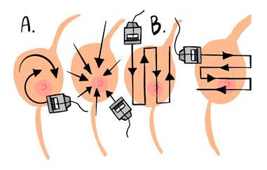
Example of different scanning patterns: a) probe movement in a radial (left panel) and anti-radial fashion (right panel); and b) demonstrates a more traverse and longitudinal scanning pattern moving in two orthogonal planes.
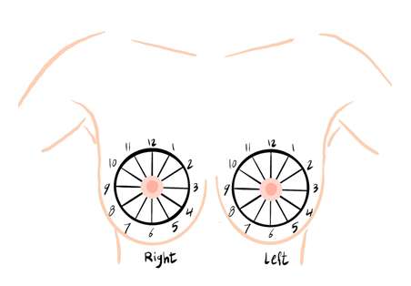
The location of breast findings on ultrasound are described by both orientation on a clock face and distance from the nipple. Each breast is described as an individual clock, with 12 o’clock at the superior breast and 6 o’clock at the inferior breast (example shown below). Of note, the clock face numbers differ between the right and left breasts due to the mirroring lateral and medial aspects (e.g. 3 o’clock is lateral in the left breast and medial in the right breast).
Breast Anatomy on Ultrasound
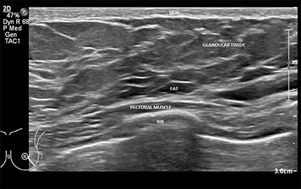
The breast can be roughly divided into three zones: the subcutaneous/premammary, mammary, and retromammary zones which can be seen depicted in the figure below.
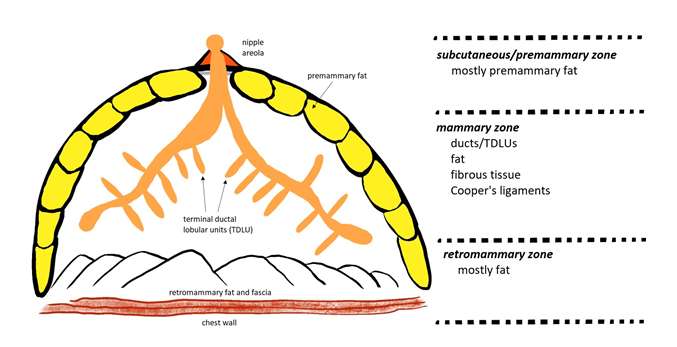
A labeled example of normal breast anatomy on ultrasound can be seen below. Of note, fat in the breast is hypoechoic, unlike ultrasound imaging of fat elsewhere in the body where is it primarily hyperechoic.
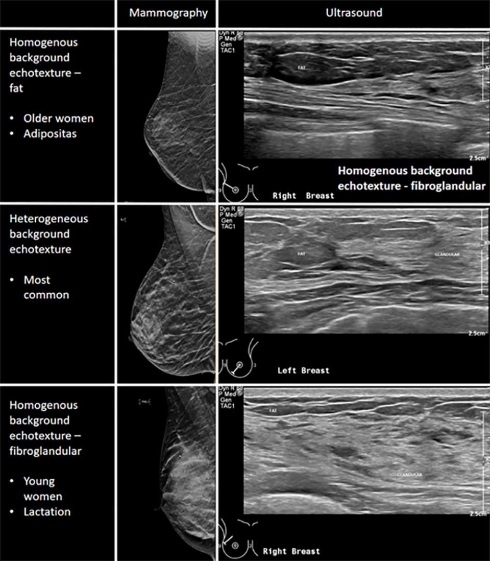
Breast Composition on Ultrasound
The breast is comprised of a mixture of fatty and fibroglandular tissue. This glandular tissue may be heterogeneously spread throughout the breast, usually more prominent within the upper outer quadrant and centrally in the breast. Glandular tissue can be affected by hormonal fluctuations, for example during the menstrual cycle, pregnancy, or lactation. Additional factors such as medications and age will affect the density of the breast tissue. Based on the Breast Imaging Reporting & Data System (BI-RADS), the lexicon to describe breast tissue composition include homogenous background echotexture – fat, homogenous background echotexture – fibroglandular, heterogeneous background echotexture.