Ultrasound: Masses
by Jonathan Lin, MD and Cheryce Fischer, MD
According to the ACR BI-RADS (Breast Imaging-Reporting and Data System) atlas, a mass is 3-dimensional lesion that occupies space. Masses on ultrasound should be seen and evaluated in three planes. There are 5 morphologic descriptors used when reporting a mass seen on ultrasound. These descriptors include: shape, orientation, margin, echo pattern and posterior features.
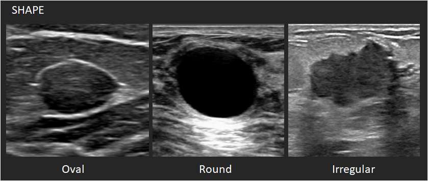
Shape:
- Oval: This shape is elliptical or egg-shaped. An oval mass may include two or three undulations.
- Round: This shape is spherical, ball-shaped, circular or globular.
- Irregular: This shape is neither round nor oval.
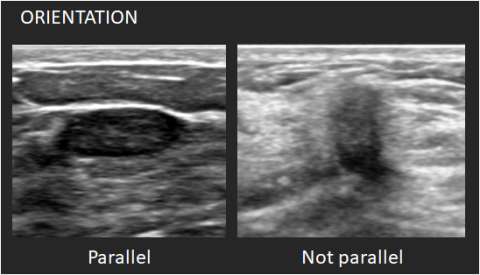
Orientation: Orientation is defined with reference to the skin line. Parallel or “wider-than-tall” orientation is often a characteristic of benign masses; however, it should not be the sole feature when assessing for likelihood of malignancy.
- Parallel: The long axis of the mass parallels the skin line.
- Not parallel: The long axis of the mass is not parallel to the skin line.
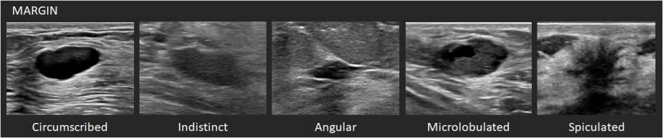
Margin: The margin is the edge or border of the lesion.
- Circumscribed: The margin is well-defined with abrupt transition between the lesion and surrounding tissue.
- Not circumscribed – If any portion or the margin is not circumscribed, the mass should be characterized as not circumscribed. The margins can be further characterized as below.
- Indistinct – No clear demarcation of the margin from the surrounding tissue.
- Angular – Some or all of the margin has sharp corners, forming acute angles.
- Microlobulated – The margin demonstrates short-cycle undulations.
- Spiculated – The margin demonstrates sharp lines radiating from the mass. This is often a sign of malignancy.
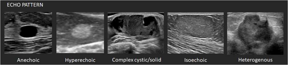
Echo pattern: The echogenicity of masses is described relative to the mammary fat.
- Anechoic – There are no internal echoes.
- Hyperechoic – There is increased echogenicity relative to fat or equal to fibroglandular tissue.
- Complex cystic/solid – There are both anechoic (fluid) and echogenic (solid) components.
- Hypoechoic – There is decreased echogenicity relative to fat.
- Isoechoic – There is the same echogenicity as subcutaneous fat.
- Heterogenous – There is a mixture of echogenic patterns within a solid mass.
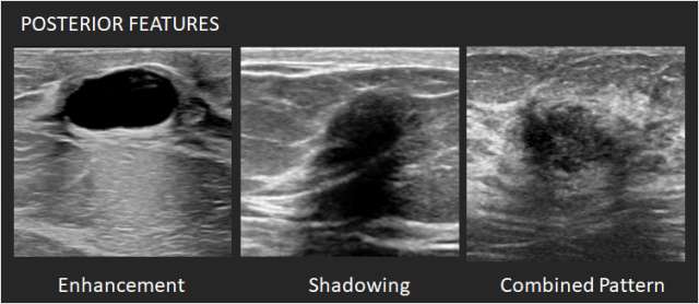
Posterior features: Posterior features represent the attenuation characteristics of a mass in respect to its acoustic transmission.
- No posterior features – Neither posterior shadowing or enhancement is seen.
- Enhancement – There is a column that is more echogenic deep to the mass. One criterion for cyst diagnosis is enhancement.
- Shadowing – There is attenuation of acoustic transmission with the area posterior to the mass appearing darker. Shadowing is associated with fibrosis, with or without an underlying carcinoma. Macrocalcifications can also cause shadowing.
- Combined pattern – There is more than one pattern of posterior attenuation.
References:
- Sickles, EA, D’Orsi CJ, Bassett LW, et al. “ACR BI-RADS® Mammography.” In: ACR BI-RADS® Atlas, Breast Imaging Reporting and Data System. Reston, VA, American College of Radiology; 2013.