Breast Asymmetry
by Bill Zhou, MD, and Anne Hoyt, MD
Asymmetry is a general term used to describe unilateral increased fibroglandular density without meeting criteria for mass, which is a three-dimensional space occupying finding usually with convex margins1. Asymmetries are common and see in 3% of mammograms2. They are nonspecific and can represent benign findings such as overlapping tissue or malignant etiologies such as invasive ductal carcinoma or invasive lobular carcinoma. In the absence of clear imaging stability, asymmetries seen on screening exams are usually recalled fora diagnosticwork up, including additional mammographic views and ultrasound if necessary.
There are four types of asymmetry according to the BI-RADS Atlas3.
Asymmetry:
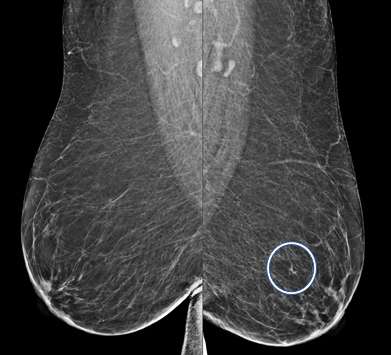
An asymmetry is a finding onlyseen on one projection3. Asymmetries that have been evaluated without suspicious features but persist are likely benign and can be managed with 6-month short-term imaging follow up.
Focal Asymmetry:
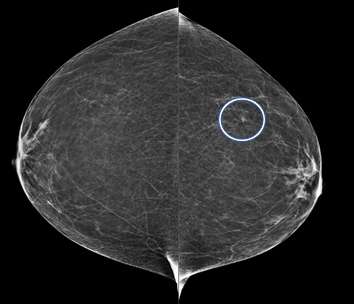
A focal asymmetry is a finding seen on two projections3. Focal asymmetries that have been evaluated without suspicious features but persist are likely benign and can be managed with 6-month short-term imaging follow up.
Developing Asymmetry:
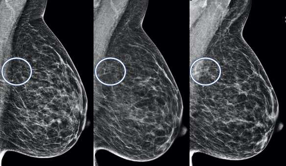
A developing asymmetry is an asymmetry that is larger or more conspicuous compared to prior exams3. Since breast tissue tends to become less dense with age, developing asymmetries are usually more concerning and may warrant additional workup such as biopsies.
Global Asymmetry:
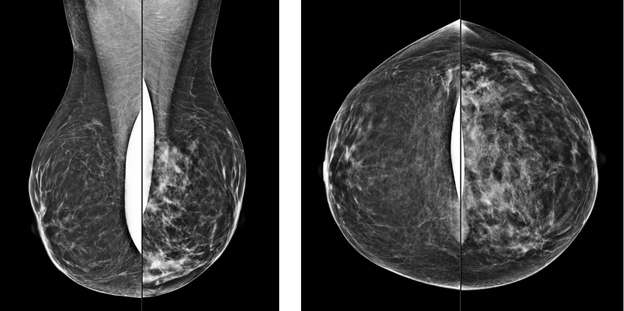
A global asymmetry is a finding seen on two projections of significantlyincreased breast tissue occupying greater than one quadrant. Global asymmetries that have been evaluated without suspicious features are most lynormal variantsand do not require additional follow up.
Common benign etiologies1:
- Overlapping fibroglandular tissue
- Anatomicvariant
- Ectopic tissue
- Post-surgical
- Hematoma
- Fat necrosis
- Radial scar
- Cysts
- Sclerosing adenosis
- Hormonal replacement
- Oral contraceptives
- Diabetic mastopathy
Common malignant etiologies1:
- Invasive ductal carcinoma
- Invasive lobular carcinoma
- Lymphoma
References:
- Samardar P, De paredes ES, Grimes MM et-al. ”Focal asymmetric densities seen at mammography: US and pathologic correlation.” Radiographics 22(1):19-33. DOI: 10.1148/radiographics.22.1.g02ja2219
- Kopans DB, Swann CA, White G, et al. “Asymmetric breast tissue.” Radiology 1989; 171:639-64. DOI: 10.1148/radiology.171.3.2541463
- D’Orsi CJ, Sickles EA, Mendelson EB, Morris EA, et al. ACR BI-RADS® Atlas 5th Edition, Breast Imaging Reporting and Data System. Reston, VA, American College of Radiology; 2013. https://www.acr.org/Clinical-Resources/Reporting-and-Data-Systems/Bi-Rads