Pediatric Diseases
Find Your Care
Our interventional neuroradiology team has paved the way for interventional procedures, creating several of the devices and techniques widely used today. Call 310-267-8761 to learn more about our services.
Pediatric Craniofacial Vascular Malformations
What are they?
They are abnormally formed vessels carrying blood or lymphatic fluid that are present at birth but may not become evident until later in life.
What are the different types of vascular malformations?
They are two different broad categories: high flow and low flow malformations. The high flow malformations are called arterio-venous malformations because they represent abnormal connections between arteries or arterioles and veins or venules which bypass the normal capillary circulation. These can also occur in the brain (brain arteriovenous malformations). In the body, arteriovenous malformations can have life-threatening bleeding or mass effect, local tissue effects, and/or cosmetic disturbance. They are often treated with embolization as described in the brain arteriovenous malformation section. The low-flow malformations are due to abnormal formation of capillaries, veins or lymphatics. Sometimes the malformations are combined such as a veno-lymphatic malformation.
How are they diagnosed?
The lesions are usually identifiable on a physical exam as a vascular lesion. The venous malformations may demonstrate enlargement with various maneuvers that in crease pressure in the veins such as bringing the lesion below the level of the heart. High-flow lesions may be associated with a pulsatile bruit on exam.
How are they evaluated?
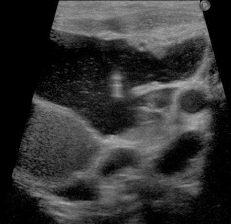
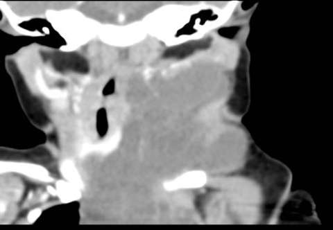
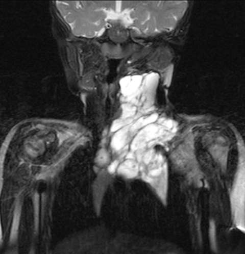
The lesions are most commonly evaluated with contrast enhanced MRI. Occasionally US and CT are used.
What are the symptoms of a vascular malformation?
The lesions are often associated with pain and cosmetic disfigurement. They can also often present with pain at the site of the malformation. They can occasionally present with bleeding, especially if they involve the lining of the nose or mouth. This bleeding can be profound with high-flow vascular malformations. In addition, the lesions involving the aero-digestive tract, such as in the throat can present with difficulty breathing and\or swallowing.
How are they treated?
A team approach is utilized in the treatment of vascular malformations. The Division of Internvational Neuroradiology (DINR) is part of a combined center, The Vascular Birthmark Clinic. The following specialties may be involved depending on the location of the lesion: dermatology, otolaygology, plastic surgery, ophthalmology, oromaxillofacial surgery, and neuro-interventional radiology. Often, minimally invasive image guided percutaneous sclerotherapy is successful for the low flow venous and veno-lymphatic malformations. In this procedure X-ray and\or high resolution ultrasound guidance is used to inject a sclerosant into the malformation to damage the cells lining the lesion so that the fluid in the lesion does not re-accumulate. Sometimes several sclerotherapy sessions may be necessary. Patients typically go home on the day of the procedure.
The high-flow vascular malformations are treated with an endovascular approach from a small puncture in the femoral artery. In this procedure, micro-catheters are used to deliver liquid embolic agents to close the abnormal arteries supplying the malformation.