Ependymoma
Find Your Care
Our expert neurosurgery team is committed to providing the finest and most comprehensive patient care. For help finding a neurosurgeon, call 310-825-5111.
About Ependymoma Brain Tumor
Ependymomas: Symptoms, Treatment and Diagnosis at UCLA
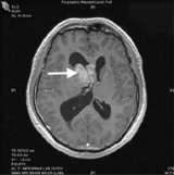
Ependymomas are tumors that arise from a thin layer of cells that line the ventricular walls and central canal of the spinal cord.
Ependymomas are a type of glioma, along with astrocytoma and oligodendroglioma. These tumors can occur anywhere in the brain or spinal cord. The most common site in the brain is at the bottom of the brain near the spinal cord. These tumors have the potential to spread through the cerebrospinal fluid (CSF) to other areas of the nervous system.
Symptoms
- Common symptoms include headache, nausea/vomiting, ataxia (difficulty with balance) or dizziness.
Diagnosis
- Magnetic resonance imaging (MRI) is the preferred diagnostic tool, although the diagnosis should be confirmed by microscopic examination of the tumor from a biopsy.
Treatment
- UCLA offers a multidisciplinary management of ependymomas, with experts in neurosurgery, radiation oncology, and neuro-oncology working closely together to assure the highest quality of care.
- Surgery
- Your UCLA neurosurgeon will determine with you how much of the tumor can and should be removed.
- If the tumor is surgically accessible, a "complete" removal is attempted.
- An actual tissue biopsy is typically required for definitive diagnosis in nearly all cases.
- Sometimes, ependymomas are best removed with the aid of intra-operative MRI, allowing the most complete, safest surgery possible.
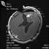
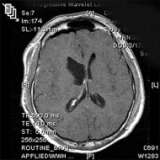 This post-operative MRI (above) confirms image-complete removal of the tumor.
 - Radiation Therapy
- Radiation therapy is administered to the tumor bed after surgical removal and if any metastases through the C
SF are found.
- Radiation therapy is administered to the tumor bed after surgical removal and if any metastases through the C
- Chemotherapy
- The UCLA Neuro-Oncology Program offers the latest in promising medical therapies available.
- Surgery
The Neuro-ICU cares for patients with all types of neurosurgical and neurological injuries, including stroke, brain hemorrhage, trauma and tumors. We work in close cooperation with your surgeon or medical doctor with whom you have had initial contact. Together with the surgeon or medical doctor, the Neuro-ICU attending physician and team members direct your family member's care while in the ICU. The Neuro-ICU team consists of the bedside nurses, nurse practitioners, physicians in specialty training (Fellows) and attending physicians. UCLA Neuro ICU Family Guide