Neurosurgery
Optic Nerve Glioma
Find Your Care
Our expert neurosurgery team is committed to providing the finest and most comprehensive patient care. For help finding a neurosurgeon, call 310-825-5111.
About Optic Nerve Glioma
General Information
- Optic nerve glioma is a type of juvenile pilocytic astrocytoma (Grade 1 astrocytoma), which occurs more commonly in children and young adults. They also have a more favorable prognosis compared with all other astrocytoma types.
- The most common site of juvenile pilocytic astrocytomas is the cerebellum.
Symptom
- The most common symptoms are headache, nausea, vomiting, poor balance, and visual complaints, depending on the tumor site. Many symptoms may be due to hydrocephalus.
Diagnosis
- Magnetic resonance imaging (MRI) is the preferred imaging technique for diagnosis.
- For surgical planning, mapping of critical speech and motor brain regions that may be adjacent to the tumor may be necessary. Such mapping is performed at the world-renowned UCLA Brain Mapping Center.
- An actual tissue biopsy is typically required for definitive diagnosis in nearly all cases. If the tumor is inaccessible to open surgery, precisely guided (stereotactic) needle biopsies can be performed.
Treatment
- Your UCLA neurosurgeon will determine with you how much of the tumor can or should be removed.
- If the tumor is surgically accessible, a "complete" removal is attempted. This can be curative.
- In cases in which the tumor cannot be removed, a conservative, non-operative approach is recommended.
- Often, juvenile pilocytic astrocytomas are best removed with the aid of intra-operative MRI, allowing the most complete, safest surgery possible.
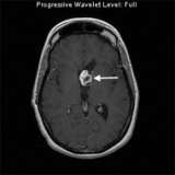
This MRI image shows a juvenile pilocytic astrocytoma in the ventricle of the left frontal lobe (arrow). This patient had a partial removal of the tumor because the tumor involved the optic nerves.