Slit Ventricle Syndrome
Find Your Care
Our expert neurosurgery team is committed to providing the finest and most comprehensive patient care. For help finding a neurosurgeon, call 310-825-5111.
About Slit Ventricle Syndrome
General Information
- Slit ventricle syndrome occurs in minority of patients who have been shunted.
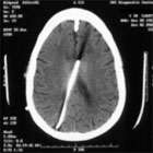
- "Slit ventricle" refers to the computed tomography (CT) or magnetic response imaging (MRI) finding of very small ("slit-like") ventricles.
- Typically, the shunt is nearly blocked but still barely flowing. Symptoms usually present years after shunt placement or revision.
- The most severe form of slit ventricle syndrome occurs in children.
- The absence of cerebrospinal fluid (CSF) within the ventricles combined with a growing brain leads to situation in which "the brain is too big for the skull."
- The intracranial pressure (brain pressure) can be very high.
- Adults can develop a milder form of slit ventricle syndrome, although most were shunted as children.
Diagnosis
- The diagnosis of slit ventricle syndrome can be difficult and the condition is often misdiagnosed or the diagnosis delayed.
- The finding of small ventricles in a shunted patient can be misinterpreted as a properly working shunt. Most patients with small ventricles (seen on CT or MRI) do not have the slit ventricle syndrome. Patients must be symptomatic.
Symptoms
- Symptoms can include headache, varying degrees of lethargy, with or without nausea, and vomiting. The symptoms may be intermittent. Over drainage is associated with positional headaches that are often relieved when lying down. Patients may be asymptomatic for prolonged periods.
Treatment
- The management of slit ventricle syndrome is difficult and challenging. In general, a neurosurgeon with expertise in the management of hydrocephalus is optimal.
- Various treatment options have been proposed, and include:
- Observation.
- Usually limited to minimally symptomatic patients
- Anti-migraine medicines.
- Shunt revision.
- Change the ventricular catheter.
- Change the shunt valve.
- Add siphon controlling device (SCD).
- Programmable valve with or without SCD.
- Converting to a lumboperitoneal shunt.
- Temporarily blocking the flow of the shunt (via "externalization" of the shunt) in order to expand the ventricles.
- This should be done with ICP monitoring due to the risk of coma.
- Many patients have aqueductal stenosis, and therefore are candidates for endoscopic third ventriculostomy (ETV).
- In some cases, a special shunt configuration draining both the ventricles and the cisterns (space around the brain) can equalize the inner and outer brain pressures, thus reducing the chance of producing slit ventricles again. This type of shunt is called a ventriculocisternoperitoneal shunt.
- Subtemporal decompression.
- This procedure is rarely performed because improvements, if any, are typically short-lived.
- Observation.
The Neuro-ICU cares for patients with all types of neurosurgical and neurological injuries, including stroke, brain hemorrhage, trauma and tumors. We work in close cooperation with your surgeon or medical doctor with whom you have had initial contact. Together with the surgeon or medical doctor, the Neuro-ICU attending physician and team members direct your family member's care while in the ICU. The Neuro-ICU team consists of the bedside nurses, nurse practitioners, physicians in specialty training (Fellows) and attending physicians. UCLA Neuro ICU Family Guide