Pineal Tumor
Find Your Care
Our expert neurosurgery team is committed to providing the finest and most comprehensive patient care. For help finding a neurosurgeon, call 310-825-5111.
About Pineal Tumors
General Information
- Tumors of the pineal region account for just 0.5 percent to 1.6 percent of human brain tumors.
- Several different types of tumors that arise in the area of the pineal gland, requiring different treatments.
- The exact diagnosis is critical for choosing the correct therapy.
- Germ cell tumors
- Account for more than half of all pineal region tumors.
- Pure germinomas account for 65 percent to 72 percent of all intracranial germ cell tumors. Germinomas have indistinct boundaries and often seed the ventricular system, which contains cerebrospinal fluid (CSF).
- The embryonal cell carcinoma, which is the least frequently reported intracranial germ cell tumor, is rarely found in its pure form and is usually highly malignant.
- Endodermal sinus tumors are rare and highly invasive.
- Teratomas can be either immature or mature and represent the expression of embryonic differentiation in germ cell tumors. These tumors are usually well formed, round or lobulated, and multicystic. They compress surrounding structures.
- Pineal parenchymal tumors
- Account for 20 percent to 30 percent of all tumors in this location.
- Two types -- pineoblastoma and pineocytoma -- differ by appearance, level of differentiation, and degree of malignancy.
- Other tumor types
- Astrocytoma
- Meningioma
- Metastatic brain tumor
- Lymphoma
- Pineal cyst
- Germ cell tumors
- Pineal tumors typically present with hydrocephalus, a build up of CSF pressure within the brain.
- A team of experts is needed for optimum therapy. UCLA experts in neurosurgery, neuropathology (microscopic diagnosis), radiation therapy, and oncology will work together to provide the best and latest therapies.
Symptoms
- Symptoms arise when the tumor invades or compresses adjacent structures. The manifestations depend on the size of the lesion and whether it is an invasive tumor.
- The involvement of the cerebral aqueduct causes hydrocephalus and increased intracranial pressure, commonly resulting in headaches, nausea, vomiting, lethargy and altered mental function.
- Involvement of the superior colliculus causes visual problems. The most common is Parinaud's syndrome, which includes inability to look upward, double vision and nystagmus, or flickering of the eyes back and forth.
- Invasion of the thalamus can cause loss of sensation to half the body, weakness of half the body and intermittent pain. Invasion of the hypothalamus may cause disturbances in body temperature and water regulation, sleepiness and weight gain.
- Additional symptoms include memory problems, lack of coordination and tremor.
Diagnosis
- Scans
- A magnetic resonance imaging (MRI) of the brain is the most important imaging study. It will show the location, size, and shape of the tumor.
- A computed tomography (CT) scan of the brain may be helpful to see if the tumor is calcified.
- Both MRI and CT scans assess for the amount of hydrocephalus.
- Biopsy
- In many cases, a biopsy is required to determine the tumor type.
- More than a dozen different types of tumor occur in this region. UCLA neurosurgeons have expertise in minimally invasive techniques (endoscopy or stereotactic needle biopsy) for obtaining biopsies.
- If the diagnosis of germinoma is suspected based on the MRI scan as well as the age and sex of the patient (young male), a biopsy may not be required prior to beginning treatment.
- Other tests
- Some tumors that occur in the pineal region have high levels of certain chemicals (Beta-HCG, AFP, and CEA), which can be detected in the cerebrospinal fluid and/or blood. If detected, a surgical biopsy may not be necessary.
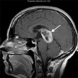
An MRI scan shows a large tumor (arrow) in the pineal region of the brain. The biopsy confirmed glioblastoma, which is rare in this part of the brain. This illustrates the importance of obtaining a tissue biopsy in most cases.
Treatment
- Surgical Removal
- The surgical removal of pineal region tumors ranks among the most difficult neurosurgical operations. Patients typically are referred to major university medical centers, like UCLA, where certain neurosurgeons have specific expertise in this type of surgery.
- Although surgery can be curative in some cases, in other cases it provides a definitive diagnosis and reduces the size of the tumor.
- Whenever possible, UCLA neurosurgeons use minimally-invasive surgical techniques
- Surgical Management of Hydrocephalus
- UCLA neurosurgeons have extensive expertise in the minimally invasive, endoscopic treatment of hydrocephalus. Typically, this can be done by an endoscopic third ventriculostomy. In other cases, a shunt may be necessary.
- Radiation therapy
- More than 70 percent of tumors are highly sensitive to radiation therapy.
- In many cases, stereotactic radiosurgery can provide excellent control of pineal tumors. UCLA uses the advanced Novalis shaped-beam stereotactic radiosurgery system, which is equally or more effective than Gamma Knife.
- Chemotherapy
- The choice is dependent on the type of tumor and the patient.
- Many tumor types need both radiation and chemotherapy.
The Neuro-ICU cares for patients with all types of neurosurgical and neurological injuries, including stroke, brain hemorrhage, trauma and tumors. We work in close cooperation with your surgeon or medical doctor with whom you have had initial contact. Together with the surgeon or medical doctor, the Neuro-ICU attending physician and team members direct your family member's care while in the ICU. The Neuro-ICU team consists of the bedside nurses, nurse practitioners, physicians in specialty training (Fellows) and attending physicians. UCLA Neuro ICU Family Guide